MakerSpace

While at the class MakerSpace, I played primarily with the Spinbots, a clever DIY, battery-powered device that draws a circular path as it spins.

I chose this activity, because there is something about automation that I find very appealing about Automation. Of knowing that once adjusted, you can have an object run a predictable sequence for all eternity (or until the battery runs out).

It was the first time I had encountered this device, yet its form revealed its function almost immediately. Assembly consisted of a base with an attached battery module and a vibrating/rotating motor, and three detachable “legs”, one containing a slot for the marker of your choice. I appreciated the minimalist design, it made wonder how it could inform other that of other devices that supervene on systematic motion.
Once unleashed, the Spinbot drew at least 100,000 circular objects. So the making was in preparing the tool, and in using it. In fact, using it required less of our participation than preparing it, the opposite of traditional mediums like brush and canvas.  From that insight, I learned that in art, the genius is sometimes in doing differently what people would least guess can be done differently.

Given more time, I would play with Makey Makey. I was able to play Beethoven fifth on bits of clay and a potato, but i don’t fully understand it.

Makerspace experience – due before class Nov. 20th:

Screen Shot 2013-11-20 at 4.33.54 PM

(I’d prefer to not show my face, thanks for understanding)

Highlight of Current Methodologies

A computer instructs the printer with a blue-print file in accordance with the desired cell type. The Organovo device consists of a stepper motor attatched to a robotic arm that orients the pump head, not at all dissimilar from the Makerbot in concept.

Two syringes are prepared. The first contains the cell-type that is the focus of the current procedure, lets say the parenchymal (functional) cells of the kidney nephron. They are usually contained in some kind of spheroid hydrogel encasing, making the entire medium look like an ink to the unaided human observer (thus the trade name BioInk). The second syringe is loaded with supplementary cell-types, structural cells, growth factors and what is known as a hydrogel. The first three components aid the growth and fusion process while the last provides a temporary 2-dimensional scaffolding.

The second syringe begins printing a hexagonal or honeycomb-patterned layer of mold. A feedback loop forms between the immediately deposited material and a triangulation sensor; this orients the tip of the syringe in real-time. In other words, the sensor allows the device to correct-course throughout the entire process.
This is important because cells are alive. Recent dermal cell bio-printing demonstrations by Wake Forest university, where a live patient with 3rd degree burns had his skin directly printed onto a wound, showcase why it is important the BioPrinter be able to accommodate for uncertainty.

A layer of parenchymal cells is deposited. At this stage, the plate may be removed and incubated, or the process may be repeated until you have several, alternating layers of cells and hydro-gel mold on top of each other.
The hydro-gel dissolves as the the extracellular matrix and the distinct cells continues to fuse inside the incubator.


The Origins of BioPrinting

Just as chemistry supervenes on physics and biology supervenes on chemistry, BioPrinting supervenes on 3d Printing. For this reason it can trace its origins to the invention of stereo-lithography by inventor Charles Hull in 1984, a technology that uses an additive layering process to render 3d digital designs as 3d physical objects. Once the photo-polymer is cured under UV light, you have a tangible version of whatever you were previously looking at on-screen.

Bio-printing’s first breakthrough occurred in 1996 when scientists Gabor Forgacs and his team began experimenting with live cells. They noticed that the cells had a tendency to organize according to their genetic programming even when outside of the animal. Furthermore the extra-cellular matrix that the cells produced, would initially be liquid and become less so as the cells matured. This window of opportunity  allowed the cells to be shaped prior to maturing and was analogous to the UV curing method utilized in materials science. That insight inspired the further development of these technologies.

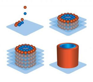
In 2000, a a human bladder was increased in size using a synthetic scaffold in a live patient. Although the cells had not been printed, they had been engineered and “taught” how to behave. When introduced to the synthetic scaffolding they arranged themselves as they would have in nature, immensely decreasing the possibility of rejection. This behavior of cells to behave as if “within the body” and arrange themselves even when grown outside the body in no particular arrangement gained prominence among scientific communities. Subsequently, research teams around the world searched for “killer” applications. Over the next decade entire organs (Hearts, kidneys, and lungs) were grown to functioning states (although not implantable) on collagen scaffolding.

heart

In 2003, a Professor from the University of Texas at Paso outfitted an inkjet printer with a cell deposition unit. This advancement, however rudimentary, proved that an additive printing process could deliver cells onto a scaffold with precision. It was only a year later that Forgacs reemerged with a method of depositing only cells, sans the scaffold, keeping them healthy enough to produce their own extra-cellular matrix and structural proteins.

http://www.wakehealth.edu/Research/WFIRM/Our-Story/Inside-the-Lab/Bioprinting.htm

The technology develops for 5 years and in 2009 Organovo, a company founded by Forgacs, introduces the Novogen MMX BioPrinter, the first market-ready version of their technology. In 2009, they print nervous tissue, followed by blood vessels in 2010, cardiac tissue in 2011, and lung tissue in 2012. Universities, notably Wake Forest, followed suit with their own versions of the technology.
Currently, they are experimenting with printing entire livers and they have reportedly done so with only a 10 day time-frame. It will still be about a decade, however, before they are of sufficient quality that they can be transplanted.

Planning Blog

11/11 – The History and Origins of BioPrinting

11/18- Highlight of Current Methodologies

12/1- BioPrinting Entrepreneurs

12/8- Bioprinting Culture: Personal Interest & the Biohacking Connection

12/14 – Future Diagnostics

12/15- How Will I Break Into the Industry?